Vitreoretinal surgery
Vitreoretinal surgery is a large family of diverse procedures involving the vitreous cavity and retina. It includes surgery for complications of cataract, retinal detachment, macula, vein thrombosis, diabetic retinopathy, and other retinal vascular disorders, ocular trauma, and intraocular infection among other things.
In all cases, these are serious retinal pathologies that can compromise a good visual recovery.


The vitreous humour is a transparent, colourless gelatinous mass that fills the space between the lens and the retina, which lines the back of the eye. This vitreous is not essential for vision and is sometimes the source of various pathologies. The retina is made up of a central part, called the macula, which is used to see details. The rest of the retinal periphery gives us the perception of the visual field.
In vitrectomy, the vitreous is removed from the eye, the retina is reapplied with intraocular gas and the tear treated with cryopexy or laser. Intraocular gas is left in the eye to ensure proper healing.
It is gradually replaced by the aqueous humour secreted by the eye. As long as the gas is present, the head should be positioned correctly and the patient should not travel to high altitudes (gas, mountains) because of the risk of expansion of the intraocular gas.
The different retinal pathologies
The CNVO treats the main retinal pathologies requiring surgery:
- Retinal detachment: uplift of the retina usually associated with a peripheral tear of the retina requiring urgent treatment.
- The epi-retinal membrane: the appearance of a small skin on the surface of the retina which causes a folding of the retina, resulting in a progressive reduction in vision and visual deformities.
- Macular hole: a hole in the centre of the retina that causes a significant visual impairment.
- Vitreoretinal traction: localized traction of the vitreous on the retina, resulting in progressive visual impairment.
Technological advances
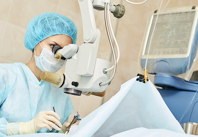
Vitreoretinalsurgery has seen many advances in recent years. The anaesthesia technique is sometimes simplified (local anaesthesia), the instruments used are increasingly fine (their diameter is 0.5 mm for the 25 gauge instruments), the visualization of the retina is improved (panoramic systems).
All of these technical improvements make it possible to carry out operations more quickly, with better post-operative comfort for the patient.

